Unlocking the Future of Medical Diagnostics: The Breakthrough Power of Time-Gated Imaging in Biomedical Applications. Discover How This Cutting-Edge Technology Is Changing the Way We Detect and Understand Disease.
- Introduction to Time-Gated Imaging: Principles and Technology
- Advantages Over Conventional Imaging Methods
- Key Applications in Biomedical Diagnostics
- Case Studies: Real-World Impact on Disease Detection
- Technical Challenges and Solutions
- Integration with Other Diagnostic Modalities
- Future Prospects and Emerging Trends
- Ethical Considerations and Regulatory Landscape
- Conclusion: The Road Ahead for Time-Gated Imaging in Medicine
- Sources & References
Introduction to Time-Gated Imaging: Principles and Technology
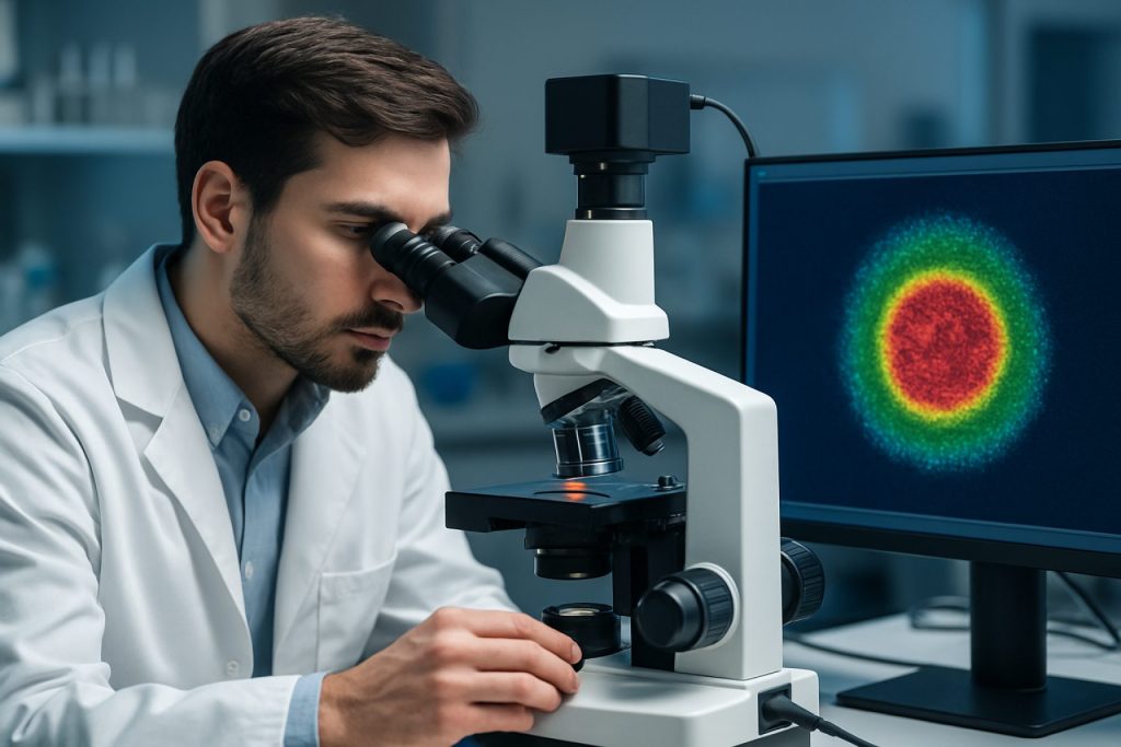
Time-gated imaging is an advanced optical technique that leverages the temporal dynamics of light emission to enhance contrast and specificity in biomedical diagnostics. Unlike conventional imaging, which collects all emitted light regardless of its origin or timing, time-gated imaging selectively captures photons within a defined time window following excitation. This approach exploits differences in fluorescence lifetimes or delayed emission properties between target signals and background autofluorescence, enabling the suppression of unwanted background and improving signal-to-noise ratios.
The core principle involves synchronizing a pulsed excitation source—such as a laser or LED—with a fast, time-resolved detector. After the excitation pulse, the detector is activated only during a specific time gate, typically nanoseconds to microseconds later, to collect photons from long-lived probes while excluding short-lived background signals. This temporal discrimination is particularly valuable in biological tissues, where endogenous autofluorescence often overlaps spectrally with exogenous labels but decays much faster. By tuning the time gate, researchers can isolate signals from probes with engineered lifetimes, such as lanthanide complexes or quantum dots, thereby achieving higher contrast and sensitivity.
Technological advancements have driven the development of time-gated imaging systems, including intensified charge-coupled device (ICCD) cameras, time-correlated single-photon counting (TCSPC) modules, and gated photomultiplier tubes (PMTs). These components enable precise control over detection timing and facilitate integration with existing microscopy platforms. The adoption of time-gated imaging in biomedical diagnostics has opened new avenues for applications such as multiplexed biomarker detection, in vivo imaging, and early disease diagnosis, as highlighted by organizations like the Nature Biomedical Engineering and the National Institute of Biomedical Imaging and Bioengineering.
Advantages Over Conventional Imaging Methods
Time-gated imaging offers several distinct advantages over conventional imaging methods in biomedical diagnostics, primarily due to its ability to selectively capture photons based on their arrival times. This temporal discrimination enables the suppression of background autofluorescence and scattered light, which are significant sources of noise in traditional continuous-wave (CW) imaging. As a result, time-gated imaging achieves higher contrast and improved signal-to-noise ratios, particularly in highly scattering biological tissues where conventional methods often struggle to distinguish weak signals from intense background fluorescence Nature Publishing Group.
Another key advantage is the enhanced depth resolution. By gating the detection window to coincide with the arrival of photons that have traveled the shortest, most direct paths, time-gated imaging can preferentially detect signals from specific tissue layers, reducing the impact of multiply scattered photons that degrade image clarity in CW techniques Optica Publishing Group. This capability is particularly valuable in applications such as fluorescence lifetime imaging (FLIM) and in vivo molecular imaging, where precise localization of signals is critical.
Furthermore, time-gated imaging facilitates multiplexed detection by distinguishing fluorophores based on their distinct fluorescence lifetimes, enabling simultaneous imaging of multiple biomarkers without spectral overlap. This multiplexing capability is challenging to achieve with conventional intensity-based imaging. Collectively, these advantages make time-gated imaging a powerful tool for improving diagnostic accuracy, sensitivity, and specificity in a wide range of biomedical applications National Center for Biotechnology Information.
Key Applications in Biomedical Diagnostics
Time-gated imaging has emerged as a transformative tool in biomedical diagnostics, offering enhanced contrast and specificity by exploiting the temporal dynamics of fluorescence and phosphorescence signals. One of its primary applications is in fluorescence lifetime imaging microscopy (FLIM), which enables the differentiation of tissue types and the identification of pathological changes based on the distinct lifetimes of endogenous and exogenous fluorophores. This capability is particularly valuable in cancer diagnostics, where time-gated imaging can distinguish malignant from healthy tissues with high sensitivity and specificity, even in the presence of strong autofluorescence background Nature Publishing Group.
Another significant application is in molecular imaging using targeted probes. Time-gated detection allows for the suppression of short-lived background signals, thereby enhancing the detection of long-lived luminescent probes such as lanthanide complexes or quantum dots. This approach is instrumental in tracking specific biomarkers, monitoring drug delivery, and visualizing cellular processes in vivo National Center for Biotechnology Information.
Additionally, time-gated imaging is increasingly used in point-of-care diagnostics, where portable devices leverage this technology to perform rapid and sensitive assays for infectious diseases, cardiac markers, and metabolic disorders. The ability to discriminate signal from noise in complex biological samples makes time-gated imaging a powerful platform for multiplexed detection and quantitative analysis in clinical settings Elsevier.
Case Studies: Real-World Impact on Disease Detection
Time-gated imaging has demonstrated significant real-world impact in the early detection and diagnosis of various diseases, particularly in oncology and infectious disease management. One notable case study involves the use of time-gated fluorescence imaging for the identification of sentinel lymph nodes in breast cancer surgery. By employing time-gated detection, clinicians were able to distinguish the fluorescence signal of targeted tracers from the intense background autofluorescence of surrounding tissues, resulting in improved accuracy and reduced false positives during intraoperative procedures. This advancement has led to more precise excision of cancerous tissue and better patient outcomes, as documented by National Cancer Institute.
Another impactful application is in the rapid diagnosis of tuberculosis (TB). Time-gated imaging has been utilized to detect Mycobacterium tuberculosis in sputum samples by differentiating the long-lived fluorescence of specific probes from the short-lived background signals. This approach has enabled faster and more reliable TB detection, even in resource-limited settings, as highlighted by World Health Organization. Additionally, time-gated imaging has been applied in the detection of amyloid plaques in Alzheimer’s disease, where it enhances the contrast of labeled biomarkers against brain tissue autofluorescence, facilitating earlier and more accurate diagnosis.
These case studies underscore the transformative potential of time-gated imaging in biomedical diagnostics, offering enhanced sensitivity, specificity, and speed in disease detection. As the technology continues to evolve, its integration into clinical workflows is expected to further improve diagnostic accuracy and patient care across a range of medical conditions.
Technical Challenges and Solutions
Time-gated imaging in biomedical diagnostics offers significant advantages in suppressing background autofluorescence and enhancing signal specificity. However, its implementation faces several technical challenges. One major hurdle is the requirement for precise synchronization between excitation sources and detection systems. Achieving nanosecond or even picosecond timing accuracy is essential, especially when distinguishing between short-lived autofluorescence and longer-lived probe emissions. This necessitates the use of advanced pulsed lasers and high-speed detectors, such as time-correlated single photon counting (TCSPC) modules, which can be costly and complex to integrate into clinical workflows (Nature Publishing Group).
Another challenge is the limited photon budget, particularly in deep tissue imaging, where scattering and absorption reduce the number of detectable photons. This can compromise image quality and sensitivity. Solutions include the development of brighter, longer-lived luminescent probes and the use of photon-efficient detection algorithms. Additionally, miniaturization and integration of time-gated imaging components into compact, user-friendly devices remain ongoing engineering challenges (Optica Publishing Group).
Recent advances address these issues through the adoption of solid-state detectors, such as single-photon avalanche diodes (SPADs), and the implementation of machine learning algorithms for noise reduction and signal extraction. Furthermore, the development of portable, fiber-based time-gated imaging systems is facilitating translation from laboratory to bedside, broadening the clinical applicability of this powerful diagnostic technique (National Center for Biotechnology Information).
Integration with Other Diagnostic Modalities
The integration of time-gated imaging with other diagnostic modalities has significantly enhanced the capabilities of biomedical diagnostics, enabling more comprehensive and accurate assessments of biological tissues. Time-gated imaging, which leverages the temporal separation of fluorescence or phosphorescence signals from background autofluorescence, can be combined with structural imaging techniques such as magnetic resonance imaging (MRI), computed tomography (CT), and ultrasound to provide both functional and anatomical information in a single diagnostic workflow. For instance, hybrid systems that merge time-gated fluorescence imaging with MRI allow clinicians to correlate molecular events with high-resolution anatomical structures, improving the localization and characterization of pathological changes Nature Biomedical Engineering.
Additionally, the combination of time-gated imaging with optical coherence tomography (OCT) or photoacoustic imaging enables the simultaneous acquisition of depth-resolved structural and molecular data, which is particularly valuable in oncology and cardiovascular diagnostics Elsevier – Medical Image Analysis. Integration with positron emission tomography (PET) or single-photon emission computed tomography (SPECT) further expands the diagnostic potential by allowing the correlation of metabolic or functional imaging with time-resolved optical signals. These multimodal approaches facilitate improved disease detection, monitoring, and therapy guidance by leveraging the strengths of each modality while compensating for their individual limitations National Center for Biotechnology Information.
Overall, the synergistic integration of time-gated imaging with other diagnostic technologies is driving the development of next-generation diagnostic platforms, offering clinicians a more holistic view of disease processes and enabling personalized medicine approaches.
Future Prospects and Emerging Trends
The future of time-gated imaging in biomedical diagnostics is poised for significant advancements, driven by innovations in photonics, detector technology, and computational analysis. One emerging trend is the integration of time-gated imaging with artificial intelligence (AI) and machine learning algorithms, which can enhance image reconstruction, automate feature extraction, and improve diagnostic accuracy. These approaches are expected to facilitate real-time analysis and interpretation of complex biological signals, making time-gated imaging more accessible and clinically relevant Nature Biomedical Engineering.
Another promising direction is the miniaturization and portability of time-gated imaging systems. Advances in compact ultrafast lasers and single-photon avalanche diode (SPAD) arrays are enabling the development of handheld or point-of-care devices, which could revolutionize diagnostics in resource-limited settings and at the patient bedside Optica. Additionally, the combination of time-gated imaging with other modalities, such as photoacoustic or multiphoton imaging, is expanding the range of detectable biomarkers and improving tissue penetration and specificity Nature Photonics.
Looking ahead, the translation of time-gated imaging from research laboratories to routine clinical practice will depend on further improvements in speed, sensitivity, and cost-effectiveness. Regulatory approval and standardization efforts are also critical for widespread adoption. As these challenges are addressed, time-gated imaging is expected to play an increasingly central role in early disease detection, intraoperative guidance, and personalized medicine.
Ethical Considerations and Regulatory Landscape
The integration of time-gated imaging technologies into biomedical diagnostics raises important ethical and regulatory considerations. As these advanced imaging modalities can provide highly sensitive and specific information about biological tissues, they often involve the collection and processing of detailed patient data. Ensuring patient privacy and data security is paramount, particularly as time-gated imaging may be combined with artificial intelligence or cloud-based analysis platforms. Compliance with data protection regulations such as the Health Insurance Portability and Accountability Act (HIPAA) in the United States and the General Data Protection Regulation (GDPR) in the European Union is essential to safeguard patient information and maintain public trust (U.S. Department of Health & Human Services, European Commission).
From a regulatory perspective, time-gated imaging devices intended for clinical use must undergo rigorous evaluation to demonstrate safety, efficacy, and reliability. Regulatory bodies such as the U.S. Food and Drug Administration (FDA) and the European Medicines Agency (EMA) require comprehensive preclinical and clinical data before granting approval for diagnostic applications (U.S. Food and Drug Administration, European Medicines Agency). Developers must also consider the ethical implications of incidental findings, informed consent, and equitable access to these technologies. Addressing potential biases in imaging algorithms and ensuring that new diagnostic tools do not exacerbate healthcare disparities are critical ethical challenges. Ongoing dialogue among researchers, clinicians, regulators, and ethicists is necessary to ensure that time-gated imaging advances patient care while upholding ethical standards and regulatory compliance.
Conclusion: The Road Ahead for Time-Gated Imaging in Medicine
Time-gated imaging has emerged as a transformative tool in biomedical diagnostics, offering unparalleled capabilities in enhancing image contrast, suppressing background autofluorescence, and enabling precise temporal resolution of biological events. As the field advances, the integration of time-gated techniques with other imaging modalities—such as multiphoton microscopy, super-resolution imaging, and machine learning-based analysis—promises to further expand its diagnostic potential. The development of novel luminescent probes, particularly those with long-lived emission and high biocompatibility, is expected to address current limitations related to sensitivity and specificity in complex biological environments (Nature Biomedical Engineering).
Looking ahead, the miniaturization and cost reduction of time-gated imaging hardware will be crucial for widespread clinical adoption. Portable and user-friendly devices could facilitate point-of-care diagnostics, intraoperative guidance, and real-time monitoring of disease progression. Moreover, regulatory approval and standardization of protocols will be essential to ensure reproducibility and reliability across diverse healthcare settings (U.S. Food & Drug Administration).
In summary, the road ahead for time-gated imaging in medicine is paved with opportunities for innovation and clinical impact. Continued interdisciplinary collaboration among physicists, chemists, engineers, and clinicians will be vital to translate laboratory advances into routine medical practice, ultimately improving patient outcomes and advancing the frontiers of biomedical diagnostics.
Sources & References
- Nature Biomedical Engineering
- National Institute of Biomedical Imaging and Bioengineering
- National Center for Biotechnology Information
- National Cancer Institute
- World Health Organization
- European Commission
- European Medicines Agency


